New publication from the Roukos lab: Breaking bad - How breaks in folded, active DNA promote leukaemia in response to cancer therapy
14-06-2019
Cancer is one of the most prevalent diseases worldwide, with the World Health Organisation estimating 18.1 million new cases in 2018. The predominant method for fighting cancer is chemotherapy - introducing a toxic substance to the body that preferentially kills cancer cells. However, chemotherapy can also cause severe complications. One of the worst is the development of a second cancer, usually leukaemia. New research published in Molecular Cell from the group of Vassilis Roukos at the Institute of Molecular Biology (IMB) in Mainz unveils how these treatment-related cancers can arise.
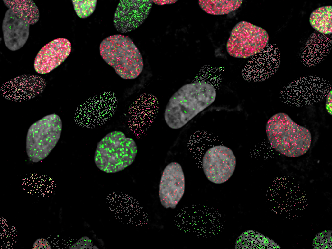
Cancers are commonly treated with anticancer drugs called topoisomerase poisons. Unfortunately, treatment with topoisomerase poisons can also cause DNA rearrangements (translocations) in healthy cells that disrupt gene regulation and lead to the development of a second cancer, usually leukaemia. However, it was unclear why these leukaemia-promoting translocations are so common after treatment with topoisomerase poisons.
In their work published in the latest issue of Molecular Cell, Dr Vassilis Roukos and his group, together with the lab of Argyris Papantonis (Center for Molecular Medicine Cologne) and the lab of Nicola Crosetto (Karolinska Institute, Stockholm), combined cutting-edge genomics and single-cell imaging methods to determine why these leukaemia-promoting translocations arise. They found that certain sites with highly active genes tend to be close to regions of DNA folding that are under more mechanical strain. This makes them susceptible to DNA breaks caused by topoisomerase poisons, producing translocations that drive leukaemia.
This finding highlights how gene activity and the arrangement of DNA within the nucleus can have a profound impact on events that trigger genomic instability to promote cancer.
For the full press release, click here.